
Past Events

In the field of mental health, the vast majority of measurements are intrinsically “messy”. This messiness stems from the difficulty in measuring ground-truth phenomena which are inaccessible. This results in off-target variations due to human interpretations, difficult-to-characterize biases, and widespread infrastructure inefficiencies. However, rather than viewing these factors solely as a hindrance, this talk explores the distinct opportunities they present to improve research robustness in research and clinical care. Drawing on concrete examples across MRI, biometrics, and clinical assessments, this presentation highlights how robust computational infrastructure can turn analytical variation into a tool for improving the generalizability of brain-behavior models. It also demonstrates how engaging the broader data science community through open data initiatives and competitions can help solve complex challenges, such as accurately detecting sleep states from wrist-worn actigraphy data. Finally, the talk illustrates how leveraging open data, software engineering, and carefully constrained Artificial Intelligence tools can transform unstructured clinical assessments into scalable, accessible tools for public good. Ultimately, this presentation outlines a principled framework for using software engineering and AI in mental health research and care in a way that can accelerate discovery and increase access without increasing the risk of harm.

Gregory Kiar, PhD, is the director of the Center for Data Analytics, Innovation, and Rigor (DAIR) at the Child Mind Institute. With a background in biomedical engineering, Greg has developed techniques to study many different forms of biosignal and health data, ranging from building turnkey tools to estimate brain connectivity maps and assess the robustness of neuroscience findings, to developing algorithms that assess personal risk from text data in journaling or electronic health records. He is a long-time advocate of open science, enthusiastic mentor, and constant seeker of interdisciplinary collaboration.

Medical imaging, including magnetic resonance imaging (MRI) and computed tomography (CT), is central to modern diagnosis and treatment planning. Although contemporary radiological AI systems can achieve fast and accurate diagnoses, most offer limited user interaction within clinical workflows. This gap hinders adoption by reducing transparency, adaptability, and trust. This talk will showcase research from our Health-X Lab on building radiological AI systems designed to collaborate with clinicians through intuitive and natural interaction channels. Specifically, I will highlight three lines of work: leveraging human visual attention during radiological reading to better align AI models with expert behavior; enabling flexible, language-based interaction with medical images; and developing interactive AI agents that support real-time, user-driven analysis. Together, these approaches illustrate how integrating visual perception, language, and interaction can transform AI from passive tools into effective clinical co-pilots, enhancing usability, interpretability, and seamless workflow integration.

Dr. Yiming Xiao is an Associated Professor in Computer Science and Software Engineering, a Concordia University Research Chair in Intuitive and Intelligent Surgical Technologies, member of the Applied AI Institute and the School of Health at Concordia, and a FRQS Junior 1 Research Scholar. He obtained his Ph.D. in Biomedical Engineering at McGill University in 2016. Soon after, he joined the PERFORM Centre as a PERFORM Postdoctoral Fellow. From 2018-2020, he was a CIHR and BrainsCAN Postdoctoral Fellow at the Robarts Research Institute of Western University. Yiming’s research combines novel techniques in medical imaging principles, computer vision, mixed reality, and deep learning to improve the efficiency and accuracy of image-based diagnosis and image-guided surgery.
Microwave breast imaging is a promising frontier in breast cancer detection. This presentation summarizes over a decade of diverse studies, from early beginnings to a prototype portable device for underserved communities. We’ve developed a Health-Canada-approved device using 24 small vector network analyzers. Ongoing efforts focus on a low-cost multistatic device. Disparities in breast cancer detection for marginalized populations highlight the need for the system’s low-cost, portable, and user-friendly solutions. A critical review emphasizes the need to improve specificity for BMI. High-contrast image reconstruction methods sometimes overshadow accuracy, a key area for investigation. The presentation focuses on radar-based reconstruction algorithms, addressing various challenges. It introduces a differential dual-breast diagnostic strategy for more accurate estimates. In conclusion, a decade of research underscores ongoing learning and integration for advancements, crucial for impacting early breast cancer detection in remote locations and low-middle income countries.
Dr. Stephen Pistorius, a tenured Professor at the University of Manitoba’s Department of Physics and Astronomy, specializes in Medical Physics. He is a Senior Scientist at the CancerCare Manitoba Research Institute and holds cross-appointments in Radiology, Biomedical Engineering, and Human Anatomy & Cell Science. A certified Medical Physicist with diverse leadership experience, he earned degrees in Physics, Radiation Physics, Medical Physics, and a Ph.D. in Physics in South Africa. Additionally, he holds a Post-Graduate Diploma in Business Management from the Edinburgh Business School, UK. Dr. Pistorius is a senior member of the IEEE and a Fellow of the Canadian Organization of Medical Physics (FCOMP). With a background in military, industry, healthcare, administration, and academia, he has held key roles and directed medical physics programs. His research, spanning image processing, reconstruction, medical device development, and diagnostic methods, includes recent work on breast microwave imaging for screening disparities in remote regions and novel PET imaging techniques. Dr. Pistorius has made significant contributions, leading national grants, authoring 300+ publications, and currently supervising twelve trainees, showcasing his commitment to advancing medical physics for improved healthcare outcomes.
EM-based medical technologies are receiving increasing interest in the global scientific community. One of the most studied applications in this field is the use of microwaves for cancer diagnosis, particularly for breast cancer detection, made possible by the significant dielectric contrast between healthy and neoplastic tissues. Several microwave imaging prototypes for breast cancer detection have been proposed, some even tested in pre-clinical settings on volunteers and patients, demonstrating reasonable target detection capabilities in dense/medium-dense tissues. In this seminar, an overview of the work done and on-going at the University of Pavia investigating the potential of millimeter (mm-) waves for early-stage breast cancer detection in fat tissues will be disclosed. In particular, the main design steps will be shown and critically discussed. In addition, the perspectives of multi-modal approaches (based on mm-waves, Ultrasounds, and elastography) to improve the cancer detection capabilities in different breast compositions will be presented.
Simona Di Meo (Member, IEEE) was born in 1992. She received the M.Sc. degree (cum laude) in electronic engineering and the Ph.D. degree in electronics and computer science from the University of Pavia, Pavia, Italy, in 2016 and 2020, respectively. She is currently a Postdoctoral Researcher with Microwave Laboratory and Bioengineering Laboratory, Department of Electrical, Computer, and Biomedical Engineering, University of Pavia. Her current research interests include innovative techniques for early breast cancer detection. She was the Co-Principal Investigator (Co-PI) with the University of Pavia funding BlueSky Research Project MULTIWAVE.
She was the Education and research cooperation committee Chair and member of the Technical Program Committee for the Mediterranean Microwave Symposium 2022, and Student Activity Chair for the European Microwave Week 2022.
She is the recipient of several awards and recognitions, including the IEEE AP-S Eugene F. Knott Memorial Pre-Doctoral Research Award, the IEEE AP-S Doctoral Research Grant, the IEEE AP-S Fellowship, the “Pavia Donna ADOS” (Association of Breast-operated Women) Award, and multiple URSI Young Scientist Awards.
Dr. Di Meo is Associate Editor of the IET Healthcare Technology Letters and Editorial Board member of the IEEE Open Journal of Engineering in Medicine and Biology.


The use of numerical exposure evaluations performed with high-resolution anatomical models has become an essential part of the safety assessment of radiofrequency (RF) fields over the last two decades, in applications from wireless communication and power transfer to wearable and implanted medical devices. This lecture reviews those applications of human anatomical models, and discusses the two important trends going forward: the first, using a wide range of anatomical models to perform in silico clinical trials and define a representative envelope of potential outcomes for the population; the second, using morphed and posed models to create a ‘digital twin’ to perform precise assessments relevant for a specific person.

Tolga Goren is Head of Customized Research at the IT’IS Foundation, which was established in 1999 through the initiative and support of the Swiss Federal Institute of Technology (ETH) Zurich, the global wireless communications industry, and several governmental agencies, and is headquartered in Zurich, Switzerland. IT’IS performs fundamental research, performs R&D services for industrial partners, and participates in the development of international safety standards. Tolga’s team performs evaluations of RF safety including, among others, active implanted medical devices according to ISO 10974 and wireless devices according to IEC 62209-1528. He studied Materials Science and Applied Mathematics at Rensselaer Polytechnic Institute in New York, and completed his doctorate at ETH Zurich in 2014.

In this lecture I will present a Functional Electrical Stimulation Therapy for reaching and grasping, which does not belong to a typical “garden variety” neuroprostheses that one can commonly find discussed in the literature. In the literature, the neuroprostheses for grasping are used to replace and substitute function, and patients are expected to depend on these devices their entire lives to reach and grasp objects. This lecture will showcase a neuroprosthetic application, better known as Functional Electrical Stimulation Therapy, which is aimed at restoring voluntary arm and hand function after severe stroke and spinal cord injury, instead of making the users depend on technology to perform reaching and grasping. In the lecture the results of two Phase II randomized control trial will be presented, which were pivotal for this technology to become a commercially viable product.

Milos R. Popovic is the Director of The KITE Research Institute at the Toronto Rehabilitation Institute – University Health Network, and a tenured Professor in the Institute of Biomaterials and Biomedical Engineering at the University of Toronto. He is a Fellow of the Canadian Academy of Engineering and the American Institute of Medical and Biological Engineering. Dr. Popovic is the co-founder and director of MyndTec, the Centre for Advancing Neurotechnological Innovation to Application (CRANIA), the CRANIA Neuromodulation Institute, and the Canadian Spinal Cord Injury Rehabilitation Association. He also founded the Rehabilitation Engineering Laboratory at KITE and held the Toronto Rehab Chair in Spinal Cord Injury Research from 2007 to 2017. He received his Ph.D. in mechanical engineering from the University of Toronto in 1996 and his Dipl. Electrical Engineer degree from the University of Belgrade in 1990. His expertise includes functional electrical stimulation, neuroprostheses, neurorehabilitation, neuromodulation, brain-machine interfaces, physiological control systems, assistive technology, robotics, modelling and control of dynamic systems, and signal processing. Dr. Popovic has received numerous awards for research, innovation, and entrepreneurship. These include the Swiss National Science Foundation Technology Transfer Award, the Engineering Medal for Research and Development, the Morris Milner Award for contributions to assistive technologies, the University of Toronto Inventor of the Year Award, the University Health Network Inventor of the Year Award, the Accessibility Innovation Showcase and Tech Pitch Competition Award, the Jonas Salk Lifetime Achievement Award, and the Engineering Medal for Entrepreneurship.

Atmospheric pollution is one of serious risk factors for human health. Especially the emission of solid aerosols, such as asbestos, carbon dust, heavy metals, etc., remains a huge problem for society. The size of particles can directly be linked to their potential for causing health problems. Particulate matter (PM10), which are particles smaller than 10 micrometers in diameter, pose the greatest problems, because they can get deep into the human body and deposit in the lungs or even infiltrate the bloodstream. To protect human health, governments have set strict limit values for particulate matter (i.e. the EU has set a limit of 40 µg/m3 of PM10 in the atmosphere). However, in order to prevent pollution it is important to monitor the amount of emitted particles directly at the source, which requires capable sensor systems.
This talk gave an overview how well radar technology may be suited for this task and showed its benefits compared to other methods. The physical relations of turbulent particle streams, electromagnetic scattering and the radar equation were discussed. Furthermore, insight was provided into the development process of the multistatic dual-frequent sensor system operating at 91.5 GHz and 150.3 GHz. Finally, measurement results obtained in various experiments covering different scenarios were presented.

Alwin Reinhardt (S’16) received the M.Sc. degree in electrical engineering and information technology from Kiel University, Germany, in 2013, where he is currently pursuing the Dr.-Ing. degree at the Chair of Microwave Engineering of Prof. Höft. His thesis („A Millimeter Wave Radar Sensor for Monitoring Solid and Liquid Aerosol Streams“) is motivated by solving the problem of measuring industrial emissions (such as particulate matter) and thus preventing atmospheric pollution. During his doctoral research Mr. Reinhardt has published over 10 peer-reviewed IEEE papers as main author. He was a recipient of the German National Academic Foundation Scholarship during his studies. At the IEEE International Microwave Symposium in Philadelphia 2018 he was awarded with the third place in the Best Student Paper Competition. He was also selected as the winner of the Innovations Competition 2018 by the German state of Schleswig-Holstein and was an invited speaker at the world’s largest industrial fair, the Hannover Messe 2019. Recently, he joined the research and development department of Rohde & Schwarz, Kiel, Germany, as a system architect.

This talk gave an overview of the development of several emerging technologies that capitalize on advances in wireless technologies to address clinically informed needs. From the high-frequency microwave range to the low-frequency kilohertz range, a contrast in the electrical properties exists between different biological tissues. These differences can be exploited in the development and application of minimally invasive electromagnetic-based medical technologies. Specifically, the design, fabrication, and the clinical testing pathway of prototypes addressing specific clinical needs, including breast cancer screening, brain bleed detection, bladder monitoring, and neutrophil monitoring during chemotherapy will be addressed. This presentation concluded with a discussion on future, unexplored avenues of research for such electromagnetic-based technologies.
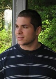
Dr. Adam Santorelli is an IRC Government of Ireland Postdoctoral Fellow in the Translational Medical Device Lab at the National University of Ireland, Galway. He studied at McGill University, Montreal, Canada, where he received his B. Eng, M. Eng, and Ph.D. in Electrical Engineering in 2010, 2012, and 2017, respectively. He has been with the Translational Medical Device Laboratory at National University of Ireland Galway since 2016, initially as a visiting researcher, and full-time as a postdoctoral researcher since 2017.
Dr. Santorelli’s current research is motivated by novel medical applications of electromagnetic and electrical engineering technologies. His work is focused on the development of compact and low-cost medical devices with the primary goal of increasing the accessibility to technology for improved diagnosis and treatment of diseases. His current work projects are focused on the development of a novel sensor to characterize and monitor changes in white blood cell count during chemotherapy, the analysis and classification of EIT data for bladder and brain trauma monitoring, and the study of the impact of various physiological processes on the dielectric properties of blood.
Dr. Santorelli has published over 50 peer-reviewed journal papers and conference proceedings. He is an active member of the Institute of Electrical and Electronics Engineers (IEEE), acting as a reviewer for several IEEE journals (TBME, TIM, TMTT, TMI, and IEEE Access) and is a part of the MyWAVE (European network for advancing Electromagnetic hyperthermic medical technologies) CA17115 COST Action.

Medical devices are increasingly investigated in the academic sector with the same rigor present in a commercial setting, thus ensuring that any technology developed has a realistic chance of translating from “research bench to patient bedside” and making a positive impact on patient care. Within the context of an aging population and an exponential growth in healthcare costs, electromagnetic (EM) therapeutic and diagnostic technologies provide an attractive solution, since they are low cost, non-ionizing, and largely non-invasive. The dielectric properties of healthy and diseased tissues are the foundation for such techniques. Despite the importance of these properties, reported values have been inconsistent, especially for key heterogeneous tissues. This inconsistency results in an unsatisfactory basis for the design and optimization of EM medical technologies, and unacceptable technical risk in the translation process. This talk discussed the state of the field, including confounders that impact the dielectric measurement of tissues, along with proposed methods for compensating for them. Needs-driven medical devices in development, including for microwave breast cancer detection, ablation of the adrenal gland to treat Conn’s syndrome, and bladder state monitoring, were also highlighted.

Dr. Emily Porter is an EU Marie-Curie Fellow and Adjunct Lecturer with the Translational Medical Device Laboratory at the National University of Ireland Galway (NUIG). She received her B.Eng., M.Eng., and Ph.D. degrees in electrical engineering from McGill University, Montreal, Canada, in 2009, 2010, and 2015 respectively. Since 2015, she has been with the Department of Electrical and Electronic Engineering at NUIG, where her current research interests include the measurement of dielectric properties of biological tissues and the development novel technologies for therapeutic and diagnostic applications of electromagnetic waves. Dr. Porter is the recipient of several prestigious national and international awards, including multiple URSI Young Scientist Awards, the IEEE Antennas and Propagation Society Doctoral Research Award, the Irish Research Council (IRC) “New Foundations” Grant, and the Royal Irish Academy (RIA) Charlemont Grant.